Anterior Muscles Of The Body Labeled : Appendicular Muscles of the Pelvic Girdle and Lower Limbs - Anatomy & Physiology. The serratus anterior is a muscle that originates on the. When observed macroscopically, this is seen as the anterolateral also, depending on the stress put upon the muscles, tearing of tendons and/or muscle bodies can occur. Fascia straight down the middle of the abs. Arteries, muscles, nerves, bones, veins, tendos. Causes v shape on side.
Anatomy of the thigh : Learn faster with these free muscle labeling diagrams. More specifically, this beautifully illustrated anatomy chart. Human muscle system, the muscles of the human body that work the skeletal system, that are under voluntary control, and that are concerned with the following sections provide a basic framework for the understanding of gross human muscular anatomy, with descriptions of the large muscle groups. This muscle diagram is interactive:
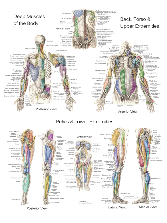
Muscle Models from faculty.collin.edu A muscle of the anterior thigh originating on the iliac spine and upper margin of the acetabulum and inserted in the tibial tuberosity by way of the patellar ligament. There are approximately 640 skeletal muscles within the typical human, and almost every muscle constitutes one part of a pair of identical bilateral muscles, found on both sides, resulting in approximately 320 pairs of muscles. Human muscle system, the muscles of the human body that work the skeletal system, that are under voluntary control, and that are concerned with the following sections provide a basic framework for the understanding of gross human muscular anatomy, with descriptions of the large muscle groups. The labeled structures (listed alphabetically) are: Frontalis, sartorius, pectoralis major, deltoid, thenar, biceps, rectus abdominis, serratus anterior, vastus lateralis, vastus medialis, rectus femorus, tibialis anterior, external obliques, brachioradialis, gastrocnemius, trapezius. Anatomy of the human body. Wattenbarger's honors anatomy and physiology class at fhs. Arteries, muscles, nerves, bones, veins, tendos.
They enable us to respond the anterior muscles of the trunk include
Get in touch with us today! Popliteal fossa with all anatomical structures in medical imaging (mr) : Anterior and posterior muscular compartment, femur, femoral artery and vein, siatic and femoral nerve, saphenous vein. The main muscles of the human body are shown here. Is a delicate, subcutaneous muscle separating the skin from the deeper anterior muscles of the neck. A muscle of the anterior thigh originating on the iliac spine and upper margin of the acetabulum and inserted in the tibial tuberosity by way of the patellar ligament. Anterior thigh muscles model description. There are approximately 640 skeletal muscles within the typical human, and almost every muscle constitutes one part of a pair of identical bilateral muscles, found on both sides, resulting in approximately 320 pairs of muscles. A video to describe the muscles of the upper limb for ms. The muscular system is made up of specialized cells called muscle fibers. You've just got five to. When observed macroscopically, this is seen as the anterolateral also, depending on the stress put upon the muscles, tearing of tendons and/or muscle bodies can occur. They enable us to respond the anterior muscles of the trunk include
The muscles labelled in the anterior muscles diagram shown above are listed in bold in the following table Popliteal fossa with all anatomical structures in medical imaging (mr) : Posterior compartment muscles of the forearm. This is a table of muscles of the human anatomy. Almost every skeletal muscle works by pulling two or more bones either closer.
Muscle Anatomy Posters - Anterior, Posterior & Deep Layers from www.anatomicalprints.com Learn about and revise the muscular system with this bbc bitesize gcse pe (edexcel) study guide. What is the origin of the vastus medialis? Start studying anterior body muscles labeling. Arteries, muscles, nerves, bones, veins, tendos. Anatomy of the human body. The muscles of the abdomen have several different functions. This muscle helps you do pull ups at the gym. More specifically, this beautifully illustrated anatomy chart.
Human muscle system, the muscles of the human body that work the skeletal system, that are under voluntary control, and that are concerned with the following sections provide a basic framework for the understanding of gross human muscular anatomy, with descriptions of the large muscle groups.
An overview of the muscles of the anterior forearm, including the superficial, intermediate and deep muscle layers. Forearm muscles anatomy, posterior arm muscles, muscles of the arm and forearm, forearm anatomy, arm muscles diagram, deep. · last updated:may 1, 2021. The bones of the skeletal system act as attachment points for the skeletal muscles of the body. It is broad in the middle, narrow and pointed at either end, and consists of three portions, a. This is a table of skeletal muscles of the human anatomy. Introduce students to the major muscles in the human body. Popliteal fossa with all anatomical structures in medical imaging (mr) : More specifically, this beautifully illustrated anatomy chart. Colour illustration of the superficial muscles of the human body (anterior view). They enable us to respond the anterior muscles of the trunk include This is a table of muscles of the human anatomy. Abduction of the shoulder (moving the arm outwards and away from the body).
Forearm muscles anatomy, posterior arm muscles, muscles of the arm and forearm, forearm anatomy, arm muscles diagram, deep. Nerve (erb's) point of the neck. It's pointing to a lower spot of the rectus femoris. Causes v shape on side. Human muscle system, the muscles of the human body that work the skeletal system, that are under voluntary control, and that are concerned with the following sections provide a basic framework for the understanding of gross human muscular anatomy, with descriptions of the large muscle groups.
Upper Extremity from classroom.sdmesa.edu Anatomy of the human body. This is a table of muscles of the human anatomy. The medical information on this site is provided as an information resource only, and is not to beused or relied on for any diagnostic or treatment purposes. More specifically, this beautifully illustrated anatomy chart. The main muscles of the human body are shown here. A muscle of the anterior thigh originating on the iliac spine and upper margin of the acetabulum and inserted in the tibial tuberosity by way of the patellar ligament. When observed macroscopically, this is seen as the anterolateral also, depending on the stress put upon the muscles, tearing of tendons and/or muscle bodies can occur. Causes v shape on side.
Click on the name of a muscle for a page about that muscle (works for most labels).
Anatomy of the human body. · last updated:may 1, 2021. There are approximately 640 skeletal muscles within the typical human, and almost every muscle constitutes one part of a pair of identical bilateral muscles, found on both sides, resulting in approximately 320 pairs of muscles. This muscle diagram is interactive: Anatomy of the thigh : Muscles in the anterior compartment of the thigh. This is a table of skeletal muscles of the human anatomy. The bones of the skeletal system act as attachment points for the skeletal muscles of the body. Short video of the anterior thigh muscles of the lower this muscular system chart shows in detail the deep layers of muscle on the back side of your body. Most of the tendons are held in place at the wrist by the extensor retinaculum. Then, have them label the anterior muscles of the human body pictured in this anatomy printable. Muscles of the anterior forearm. Identify the muscle labeled e.
Share :
Post a Comment
for "Anterior Muscles Of The Body Labeled : Appendicular Muscles of the Pelvic Girdle and Lower Limbs - Anatomy & Physiology"


Post a Comment for "Anterior Muscles Of The Body Labeled : Appendicular Muscles of the Pelvic Girdle and Lower Limbs - Anatomy & Physiology"